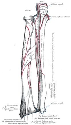
Body of ulna
| Body of ulna | |
|---|---|
 Bones of left forearm. Anterior aspect. | |
 cross-section showing the borders and surfaces of ulna and radius | |
| Details | |
| Identifiers | |
| Latin | corpus ulnae |
| TA | A02.4.06.007 |
| FMA | 33760 |
The body of the ulna at its upper part is prismatic in form, and curved so as to be convex behind and lateralward; its central part is straight; its lower part is rounded, smooth, and bent a little lateralward. It tapers gradually from above downward, and has three borders and three surfaces.
Borders
The volar border (margo volaris; anterior border) begins above at the prominent medial angle of the coronoid process, and ends below in front of the styloid process. Its upper part, well-defined, and its middle portion, smooth and rounded, give origin to the flexor digitorum profundus; its lower fourth serves for the origin of the pronator quadratus. This border separates the volar from the medial surface.
The dorsal border (margo dorsalis; posterior border) begins above at the apex of the triangular subcutaneous surface at the back part of the olecranon, and ends below at the back of the styloid process; it is well-marked in the upper three-fourths, and gives attachment to an aponeurosis which affords a common origin to the flexor carpi ulnaris, the extensor carpi ulnaris, and the flexor digitorum profundus; its lower fourth is smooth and rounded. This border separates the medial from the dorsal surface.
The interosseous crest (crista interossea; external or interosseous border) begins above by the union of two lines, which converge from the extremities of the radial notch and enclose between them a triangular space for the origin of part of the Supinator; it ends below at the head of the ulna. Its upper part is sharp, its lower fourth smooth and rounded. This crest gives attachment to the interosseous membrane, and separates the volar from the dorsal surface.
Surfaces
The volar surface (facies volaris; anterior surface), much broader above than below, is concave in its upper three-fourths, and gives origin to the flexor digitorum profundus; its lower fourth, also concave, is covered by the pronator quadratus. The lower fourth is separated from the remaining portion by a ridge, directed obliquely downward and medialward, which marks the extent of origin of the pronator quadratus. At the junction of the upper with the middle third of the bone is the nutrient canal, directed obliquely upward.
The dorsal surface (facies dorsalis; posterior surface) directed backward and lateralward, is broad and concave above; convex and somewhat narrower in the middle; narrow, smooth, and rounded below. On its upper part is an oblique ridge, which runs from the dorsal end of the radial notch, downward to the dorsal border; the triangular surface above this ridge receives the insertion of the Anconæus, while the upper part of the ridge affords attachment to the supinator. Below this the surface is subdivided by a longitudinal ridge, sometimes called the perpendicular line, into two parts: the medial part is smooth, and covered by the extensor carpi ulnaris; the lateral portion, wider and rougher, gives origin from above downward to the Supinator, the abductor pollicis longus, the extensor pollicis longus, and the extensor indicis proprius.
The medial surface (facies medialis; internal surface) is broad and concave above, narrow and convex below. Its upper three-fourths give origin to the Flexor digitorum profundus; its lower fourth is subcutaneous.
Additional images
-

Bones of left forearm. Posterior aspect.
References
This article incorporates text in the public domain from the 20th edition of Gray's Anatomy (1918)