Posterior column
| Posterior column | |
|---|---|
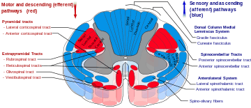
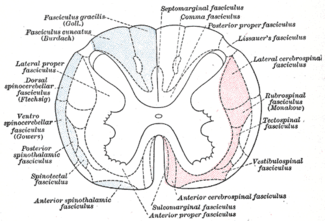
 Cross-section of the spinal cord (posterior column is labeled as dorsal column) | |
 The posterior column is made up of the gracile fasciculus and cuneate fasciculus | |
The posterior column (dorsal column) refers to the area of white matter in the middle to posterior side of the spinal cord. It is made up of the gracile fasciculus and the cuneate fasciculus and itself is part of the posterior funiculus. It is part of an ascending pathway that is important for well-localized fine touch and conscious proprioception called the posterior column-medial lemniscus pathway.[1]
Joint capsules, tactile and pressure receptors send a signal through the posterior root ganglia up through the gracile fasciculus for lower body sensory impulses and the cuneate fasciculus for upper body impulses. Once the gracile fasciculus reaches the gracile nucleus, and the cuneate fasciculus reaches the cuneate nucleus in the lower medulla oblongata, they begin to cross over as the internal arcuate fibers. Upon reaching the opposite side, they become the medial lemniscus, which is the second part of the posterior column–medial lemniscus pathway.
Lesions in this pathway can diminish or completely abolish tactile sensations and movement or position sense below the lesion.
References
- ↑ Luria, V; Laufer, E (Jul 2, 2007). "Lateral motor column axons execute a ternary trajectory choice between limb and body tissues.". Neural development. 2: 13. doi:10.1186/1749-8104-2-13. PMID 17605791.
- "The Nervous System: Sensory and Motor Tracts of the Spinal Cord" (PDF). Napa Valley College / Southeast Community College Lincoln, Nebraska. Retrieved 19 May 2013.