Pterygoid canal
| Pterygoid canal | |
|---|---|
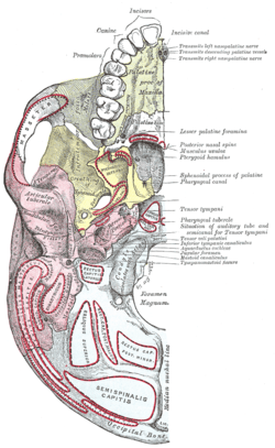
 Base of skull. Inferior surface. (Sphenoid is yellow.) | |
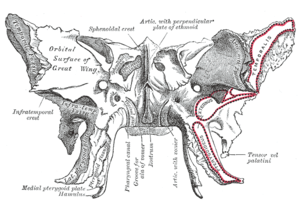
 Sphenoid bone. Anterior and inferior surfaces. (Pterygoid c. labeled at center left.) | |
| Details | |
| Artery | artery of the pterygoid canal |
| Nerve | nerve of pterygoid canal |
| Identifiers | |
| Latin | Canalis pterygoideus |
| TA | A02.1.05.053 |
| FMA | 54756 |
The pterygoid canal (also vidian canal) is a passage in the skull leading from just anterior to the foramen lacerum in the middle cranial fossa to the pterygopalatine fossa.
Structure
The pterygoid canal runs through the medial pterygoid plate of the sphenoid bone to the back wall of the pterygopalatine fossa.
Contents
It transmits the nerve of pterygoid canal, artery of the pterygoid canal and vein of the pterygoid canal.
These three are also known as the Vidian Nerve, Vidian Artery and Vidian Vein.
Additional images
 Medial wall of left orbit.
External links
- Anatomy figure: 22:4b-08 at Human Anatomy Online, SUNY Downstate Medical Center
- cranialnerves at The Anatomy Lesson by Wesley Norman (Georgetown University) (VII)
This article is issued from Wikipedia - version of the 6/9/2015. The text is available under the Creative Commons Attribution/Share Alike but additional terms may apply for the media files.