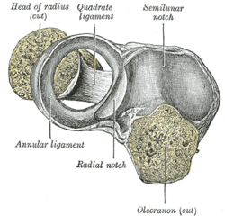
Annular ligament of radius
| Annular ligament | |
|---|---|
 Capsule of elbow-joint (distended). Anterior aspect. | |
 Annular ligament of radius, from above. The head of the radius has been sawn off and the bone dislodged from the ligament. | |
| Details | |
| Identifiers | |
| Latin | ligamentum anulare radii |
| Greek | δακτυλιοειδής σύνδεσμος |
| TA | A03.5.09.007 |
| FMA | 38872 |
The anular ligament (orbicular ligament) is a strong band of fibers that encircles the head of the radius, and retains it in contact with the radial notch of the ulna.[1]
Per Terminologia Anatomica, the spelling is "anular",[2] but the spelling "annular" is frequently encountered.
Anatomy
The anular ligament is attached by both its ends to the anterior and posterior margins of the radial notch of the ulna, together with which it forms the articular surface that surrounds the head and neck of the radius. The ligament is strong and well defined, yet its flexibility permits the slightly oval head of the radius to rotate freely during pronation and supination.[3]
The head of the radius is wider than the bone's neck, and, because the anular ligament embraces both, the radial head is "trapped" inside the ligament which thus acts to prevent distal displacement of the radius.[3]
Superiorly, the ligament is supported by attachments to the radial collateral ligament and the fibrous capsule of the elbow joint. Inferiorly, a few fibres attached to the neck of the radius support a fold of the synovial membrane without interfering with the movements at the joint.[3]
The fibrocartilage on the upper part of the ligament is continuous with the hyaline cartilage of the radial notch. At the posterior attachment the ligament widens to reach above and below the radial notch.[3]
A thickened band which extends from the inferior border of the anular ligament below the radial notch to the neck of the radius is known as the quadrate ligament.[1]
Clinical significance
Children who have not finished fusing their proximal radial epiphyseal plate may suffer dislocations of this joint. This frequently happens when parents sharply jerk their children by their arms, e.g. the act of grabbing a child away from traffic.
Additional images
|
References
This article incorporates text in the public domain from the 20th edition of Gray's Anatomy (1918)
- 1 2 Gray's Anatomy (1918), see infobox
- ↑ Federative Committee on Anatomical Terminology (1998). Terminologia anatomica: international anatomical terminology. Thieme. ISBN 978-3-13-114361-7. Retrieved 9 February 2011.
- 1 2 3 4 Palastanga, Nigel; Soames, Roger (2012). Anatomy and Human Movement: Structure and Function (6th ed.). Elsevier. p. 141. ISBN 9780702040535.
External links
- Anatomy figure: 10:5a-02 at Human Anatomy Online, SUNY Downstate Medical Center


.jpg)