Gömöri trichrome stain

Dilated peri-tubular capillaries filled with sickled RBCs, original Gomori's trichrome stain, × 400. Kanodia et al. Diagnostic Pathology 2008.[1]
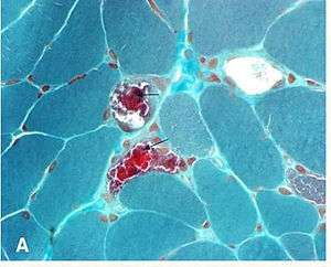
The "ragged red fibers" in MELAS syndrome are visible under modified Gomori stain.
Gömöri trichrome stain is a stain used on muscle tissue.[2][3]
It can be used to test for certain forms of mitochondrial myopathy.
It is named for George Gömöri, who developed it in 1950.[4]
References
- ↑ Kanodia KV, Vanikar AV, Goplani KR, Gupta SB, Trivedi HL (2008). "Sickle cell nephropathy with diffuse proliferative lupus nephritis: a case report". Diagn Pathol. 3: 9. doi:10.1186/1746-1596-3-9. PMC 2275218
 . PMID 18307766.
 - ↑ "GOMORI TRICHROME". Retrieved 2009-04-06.
- ↑ "Histology Lab: GOMORI TRICHROME". Retrieved 2009-04-06.
- ↑ GOMORI, G. - A rapid one-step trichrome stain. Am. J. Clin. Path. 20: 661-664, 1950.
External links
![]() Media related to Gömöri trichrome stain at Wikimedia Commons
Media related to Gömöri trichrome stain at Wikimedia Commons
This article is issued from Wikipedia - version of the 5/26/2016. The text is available under the Creative Commons Attribution/Share Alike but additional terms may apply for the media files.